Claire McKellar
Axons vs. dendrites
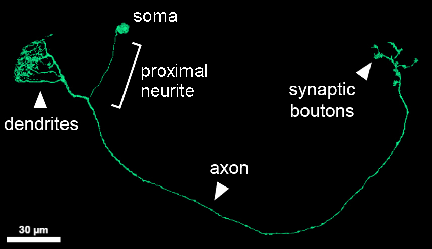
Most fly neurons are unipolar, meaning that only one neurite comes off the cell body (soma). That “proximal neurite” usually projects for some distance before branching. The location where it joins up with a thicker neurite is often (but not always) found in between two arbors: a closer one that is more dendritic in nature (finer, hairier branches, more likely to receive synaptic input than send it out), and a farther arbor that is more axonal in nature (often with the thick blobs of synaptic output terminals (boutons), and fewer fine twigs). NOTE: although these characteristics can help you orient yourself in a neuron, dendrites and axons aren’t as easy to define in flies as they are in vertebrate neurons, since there can be both input and output sites in the same arbor region.
Backbone vs. twigs
Fly neurites can be divided into larger backbones and smaller twigs, differentiated by the presence or absence of microtubules, respectively (Schneider-Mizell et al. 2016). (Microtubules are very skinny tubes running through cells.) Although backbones and twigs are technically defined by the presence or absence of microtubules, actually the absence of microtubules does not prove that something is a twig, since microtubules aren’t always visible. There are even rare exceptions where a microtubule can be seen in a small twig-like branch. Both axons and dendrites have backbones and twigs.
Twigs can be smaller than 40nm in diameter, so can get lost in between 40nm slices. This makes them difficult for the AI and for human tracers to follow, generating orphan fragments. We currently recommend ignoring orphan twigs, since it can be difficult to find their correct continuation, and future AI versions may automatically segment more twigs.
Branching
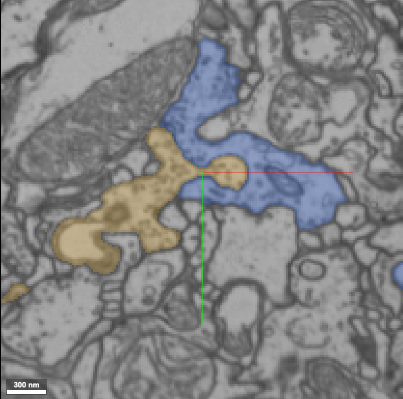
Most neurites diverge once they branch, like a tree. If you see a branch that comes off in the wrong direction, that might be a sign of an error that needs to be proofread.
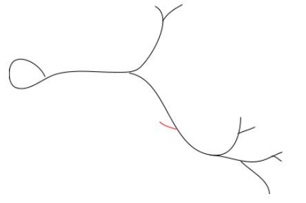
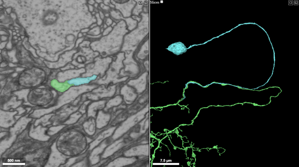
As a general rule, fly neurons don’t form loops; after two branches diverge from each other they should not merge with each other again. Occasionally, two neurites from the same neuron may appear to have rejoined, but actually just run alongside one another; this is called “auto-fasciculation”. It is rare except in the proximal neurite, which can often be found running alongside the backbone that it joins up with. This is not an error.
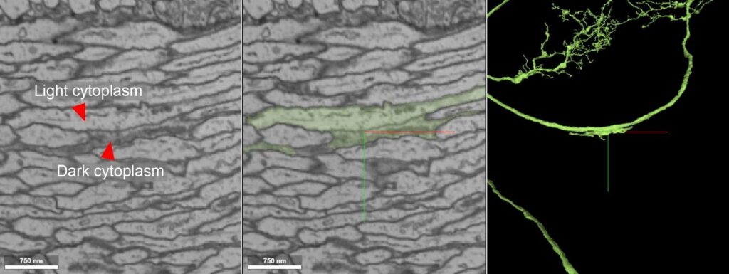
Cytoplasm
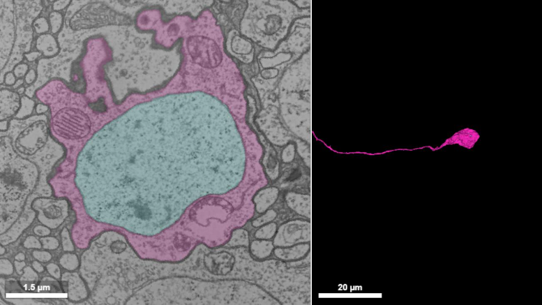
There are different types of cytoplasm. Glia are often darker than neurons, but light glial cytoplasm also exists, and dark neurons also exist (particularly certain sensory neurons).
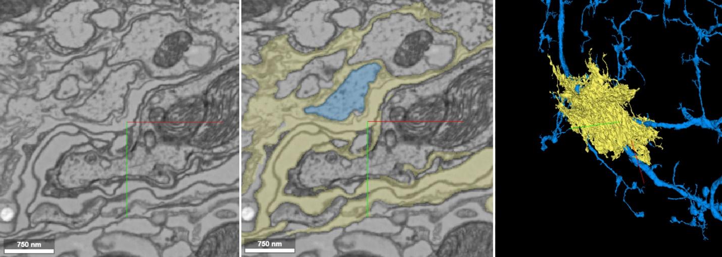
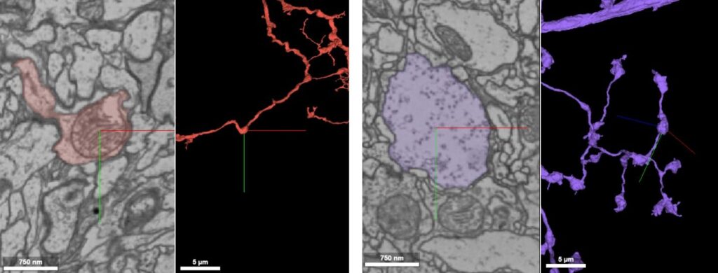
Glia (see also Cytoplasm)
Whereas neurons are roughly tubular, glia often wind between neurons, similar to how your gums form points between your teeth. Glia look a bit like seaweed in 3D, denser and thornier than neurons. They can have dark or light cytoplasm (see Cytoplasm). Glia can show accidental mergers to several other cells, and are hard to proofread. Glia frequently surround bundles of neurons as a dark boundary, sometimes making it hard to see neuron boundaries.
Membranes
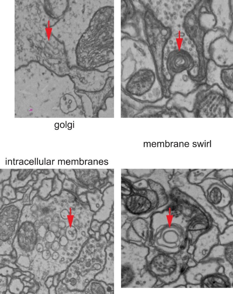
Besides the outer membrane surrounding cells, certain membranes can also be found inside cells. One examples is that cells can invaginate into each other.
There is also membrane surrounding intracellular organelles like Golgi, vesicles, mitochondria, etc. Membranes can also form swirls. (Images: Kathi Eichler.)
Microtubules and endoplasmic reticulum
Microtubules are long tubes, a little smaller in diameter than vesicles, which are present in backbones. Microtubules are not always visible, but when they are, they’re a good guide for following a neuron. Endoplasmic reticulum (ER) is also rather tube-like, slightly bigger and more variable than microtubules; it can pouch out and then become skinny again. It can also be useful for following particular neurons where it appears.
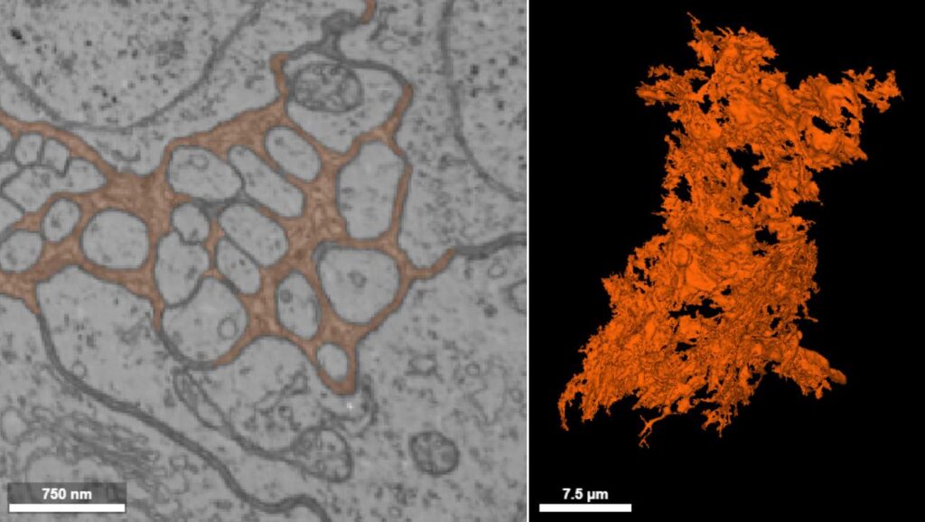
Mitochondria
Mitochondria are large intracellular organelles that produce energy for the cell. They can be round or oblong. They contain folds of membrane (cisternae) that are not always visible, or can appear to be breaking down, leaving fragments or clear spaces.

Mitochondria can cause bulges in neurites that can be confused with the bulges of synaptic boutons (although mitochondria can also be found within boutons).
Nuclei
Nuclei are occasionally segmented separately from the rest of the cell body by the AI, and have to be merged when proofreading.
Have something else you’d like to add to this glossary? Let us know!