Fly cells look a bit different from the mouse cells of EyeWire’s e2198, or even the zebrafish cells Mystics are used to working with. We can’t predict exactly what the correct morphology will be for every cell you get, but we can certainly offer a point of reference.
Let’s go over a few cells that come from the optical column, the area of the fly brain we are reconstructing. Some examples do not have completed edits, so you can see the work that still needs to be done.
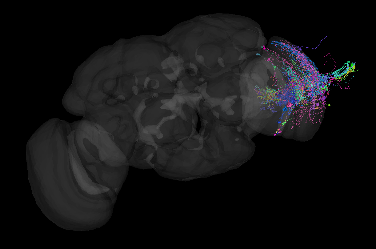
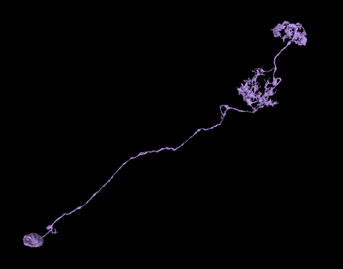
Type 1 – “Coral Fractal”
Here we see one cell type from the optical column. This cell has a somewhat dense dendritic arbor that spreads out in a single layer. Interestingly, some FlyWire cells have dendritic arbors that seem to also share axonal properties. This cell has no separate axon.
Separated segments make up this cell. It has 3 missing segments that would need to be merged before marking the cell complete. Not too many! That is typical. You don’t need to check each nub before declaring the cell complete.
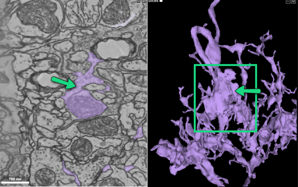
There is also a merger on this cell, highlighted in green. The merger’s morphology and direction do not match the rest of the cell, and will need to be removed.
Click here to view the cell gallery for this cell type. Please note that cells have not been fully proofread and many will include mergers or missing sections.
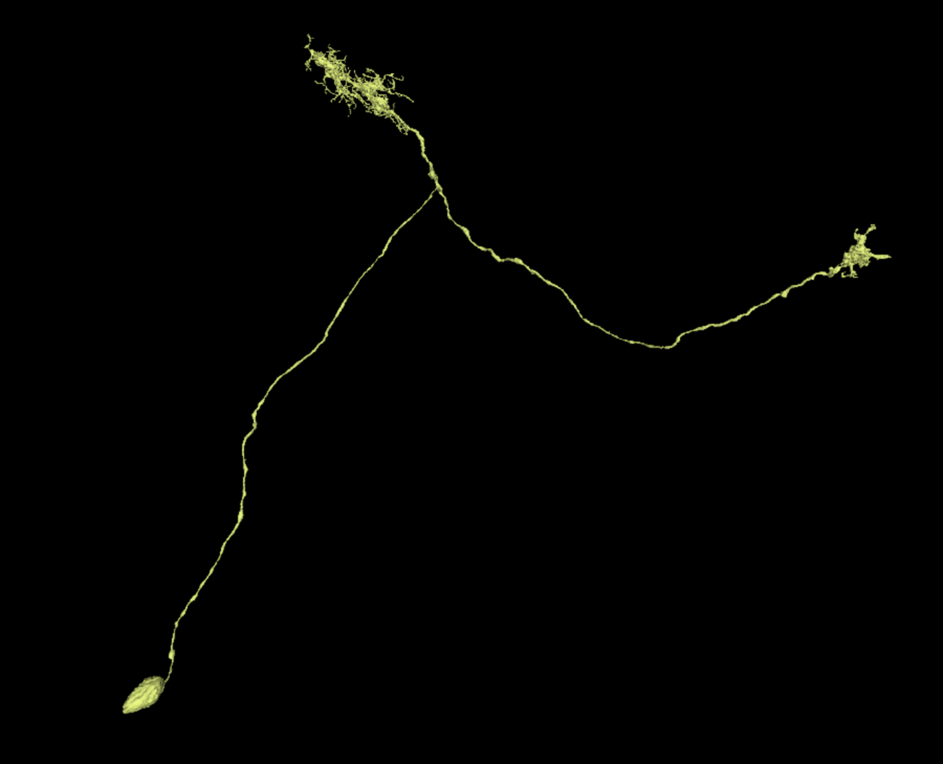
Type 2 – “Double Dendrites”
Here is another cell type from the optical column. This cell has a more “classic” configuration, in the sense that it has a separated axon (flowing left) and dendrites (going to the right). It has two small dendritic clusters.
Although it can be difficult to tell the difference between the axon and the dendrites in cells with such small dendritic clusters, the dendrites tend to be more “spiky” while the axon tends to terminate in an area that is a bit more “blobby.” Sure, those aren’t exactly scientific distinctions, but they may help you orient yourself when examining a cell.
As you can see, this cell only required 1 merge to complete. If you were proofreading this cell you would want to check over any thicker branches to see if they need extension, but you generally do not need to check each tiny twig to make sure it is finished as we are training AI to take over this task. Look at the generally morphology as a whole, and don’t sweat the tiny stuff!
Click here to view the cell gallery for this cell type.
Type 3 – “Single Shoot”
This cell has a similar morphology to the previous example, but rather than the axon and dendrites splitting off from each other, the cell moves in just one direction.
Traveling up from the soma, we first run into a dendritic cluster, and end with the axonal terminals at the top. Once again, the dendrites have a more “spiky” look, while the axon end is more “blobby.”
This cell contains a small merger on the backbone, but otherwise looks good!
Click here to view the cell gallery for this cell type.
Type 4 – “Polarized”
These t-shaped cells are similar to the “double dendrite” type, but they only have one dendritic cluster instead of 2. Remember “spiky” vs “blobby” to keep your ends straight! Can you guess which is which?
Hover here for the answer!
Click here to view the cell gallery for this cell type.
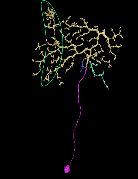
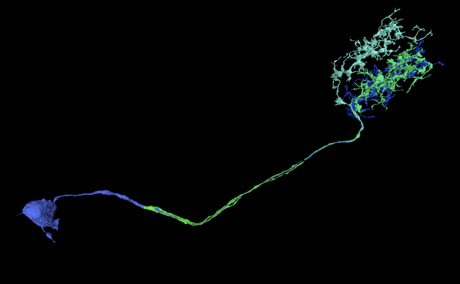
Type 5 – “Kelp”
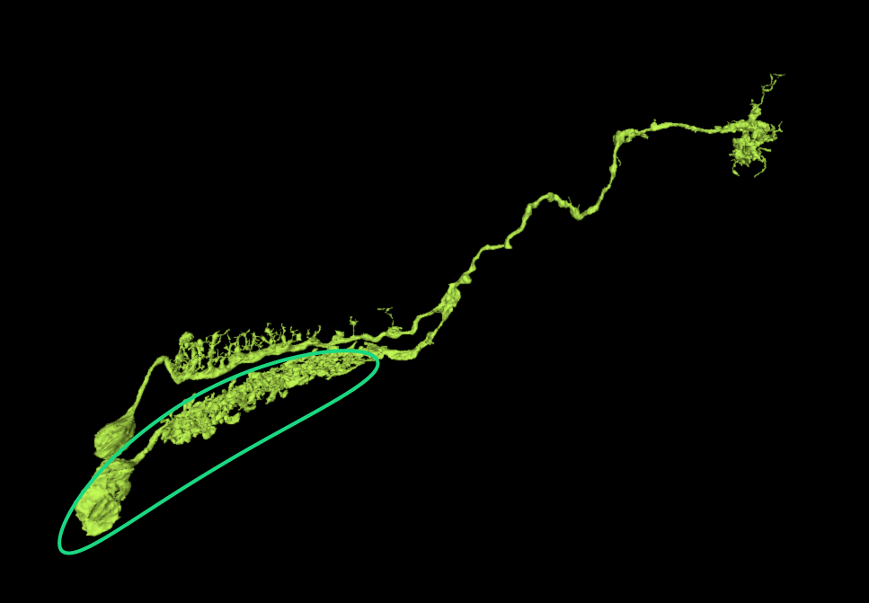
No, this cell type doesn’t have 2 cell bodies! Highlighted with the green circle you can see a merger branch that will need to be removed before this cell can be completed.
But ignoring the merger, let’s also take a look at this cell’s morphology. We start at the soma, and then move through a spiny dendritic column. We then move all the way up the cell, until we get to the end where the axon terminates. It looks a bit like a stalk of giant kelp!
Click here to view the cell gallery for this cell type.
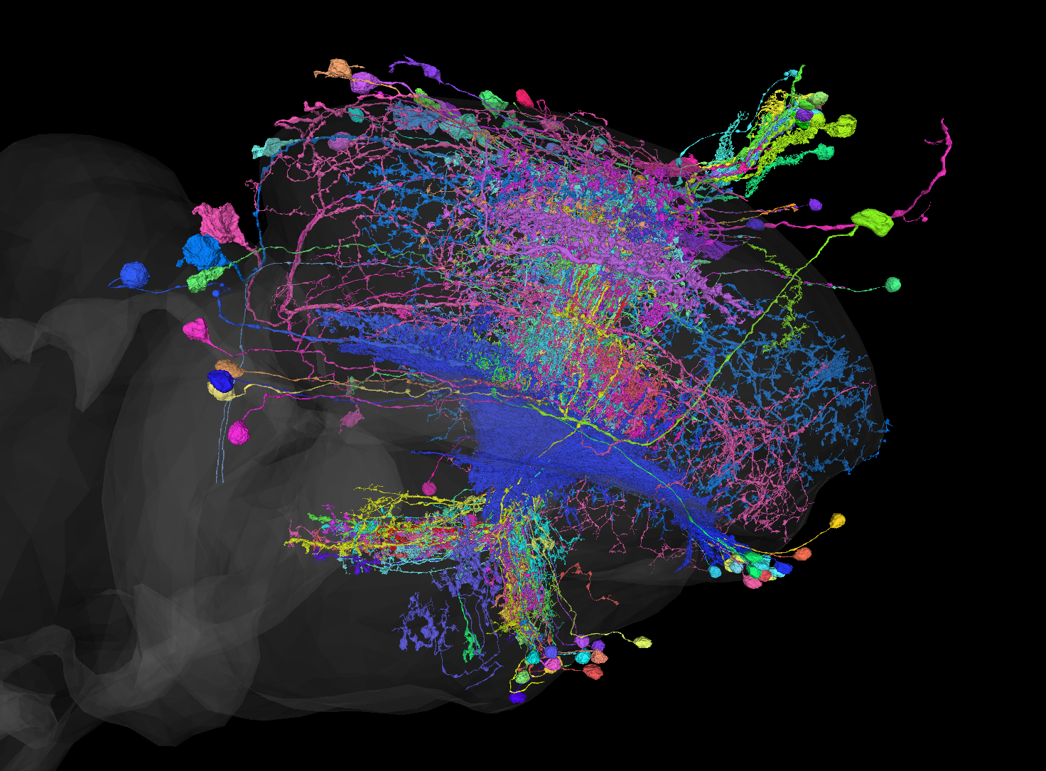
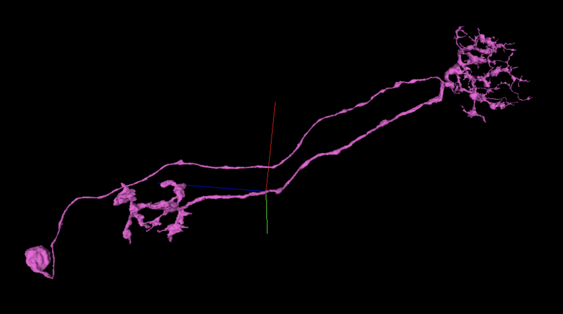
Type 6 – “Bouquet”
This is a somewhat rare, but quite interesting cell type! These cells begin normally with a single tract shooting out from the soma. However, they soon splinter into multiple tracts that continue running parallel to each other. Each one ends in its own dendritic arbor.
They look a bit like bouquets of flowers. Fascinating!
Click here to view the cell gallery for this cell type.
Type 7 – “U-Turn”
Nope that’s not a merger! This cell type heads in one direction and then decides to turn around and head home again. It features a dendritic cluster at the point furthest from the soma, and the terminal axon cluster at the branch’s end.
One response to “FlyWire Help: What will my cell look like?”
[…] Many cells, but not all, have axons. There are a fair number of FlyWire cells that do not have an axon. In these cells, the dendritic arbor has properties of both dendrites and axons. See for example, Cell Type 1 in the “What will my cell look like?” blog post. […]