If your eyes were as big as a fly’s relative to your body size, how big would each of your eyes be? Hellooooo anime eyes. Each of your irises (colorful part of the eye) would be nearly as long as your hand, about 1 cm (6 in) across.
Much of a fly’s cognitive processing power is dedicated to vision. Understanding circuits underlying vision may uncover secrets of sensory processing in other intelligent animals, such as humans.
The fly sees through hundreds of tiny eyes. Each one looks a little like the eye of Sauron. Seriously. Except in stead of the search for power, the top is a lens that focuses photons (light) into a couple different types of photoreceptors. Around the edge are photoreceptors that (we think) respond to light / dark and in the center are receptors for what eventually becomes motion sensitivity. These cells relay information about vision into cells with distinctive columnar organization. These parallel processing units relay all the visual information from a fly’s eyes to it’s brain. How do they work? We have no clue. Ok to be fair, we have a lot of clues. But we don’t *actually* know, not even enough to answer definitively what these cells do or how they do it. Mapping these regions of brain will likely unlock many new secrets of sight. Let’s check out the contenders for disrupting neuroscience.
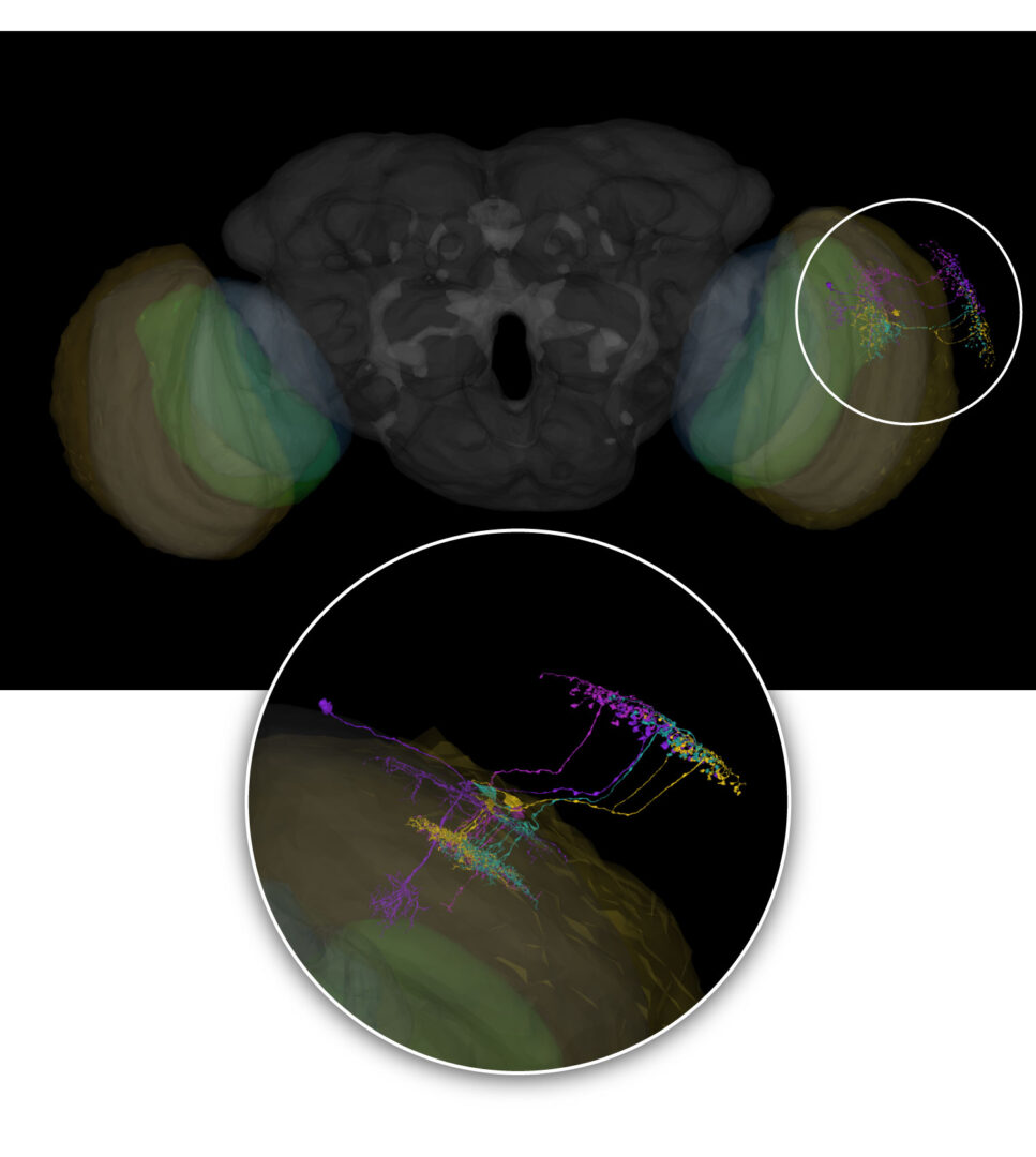
Here are the optic lobes relative to a whole fly brain. FlyWire link to brain with optic lobe regions highlighted as below. Brain regions in fly are called neuropil.
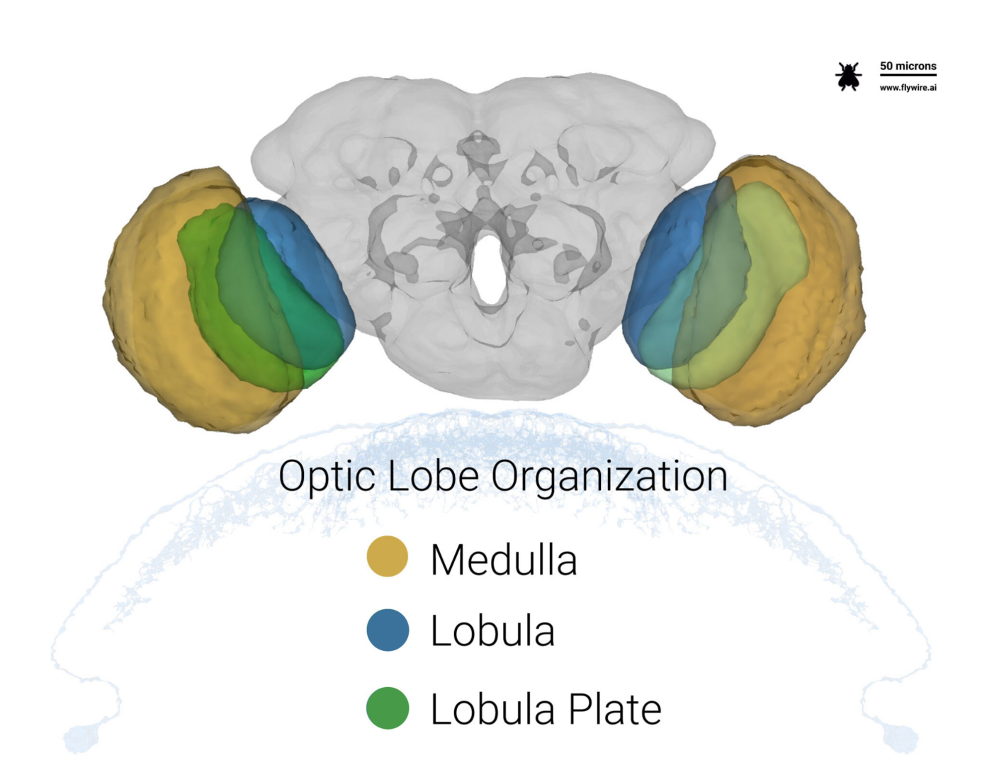
The fly’s eyes sit on top of the yellow Medulla in a region called the Lamina that isn’t shown in 3D. Our neurons from the optic column reach into the brain, passing along their visual wares to cells inside.
Let’s start from the top and work our way in.
Centrifugal Cells
FlyWire link to centrifugal cells.
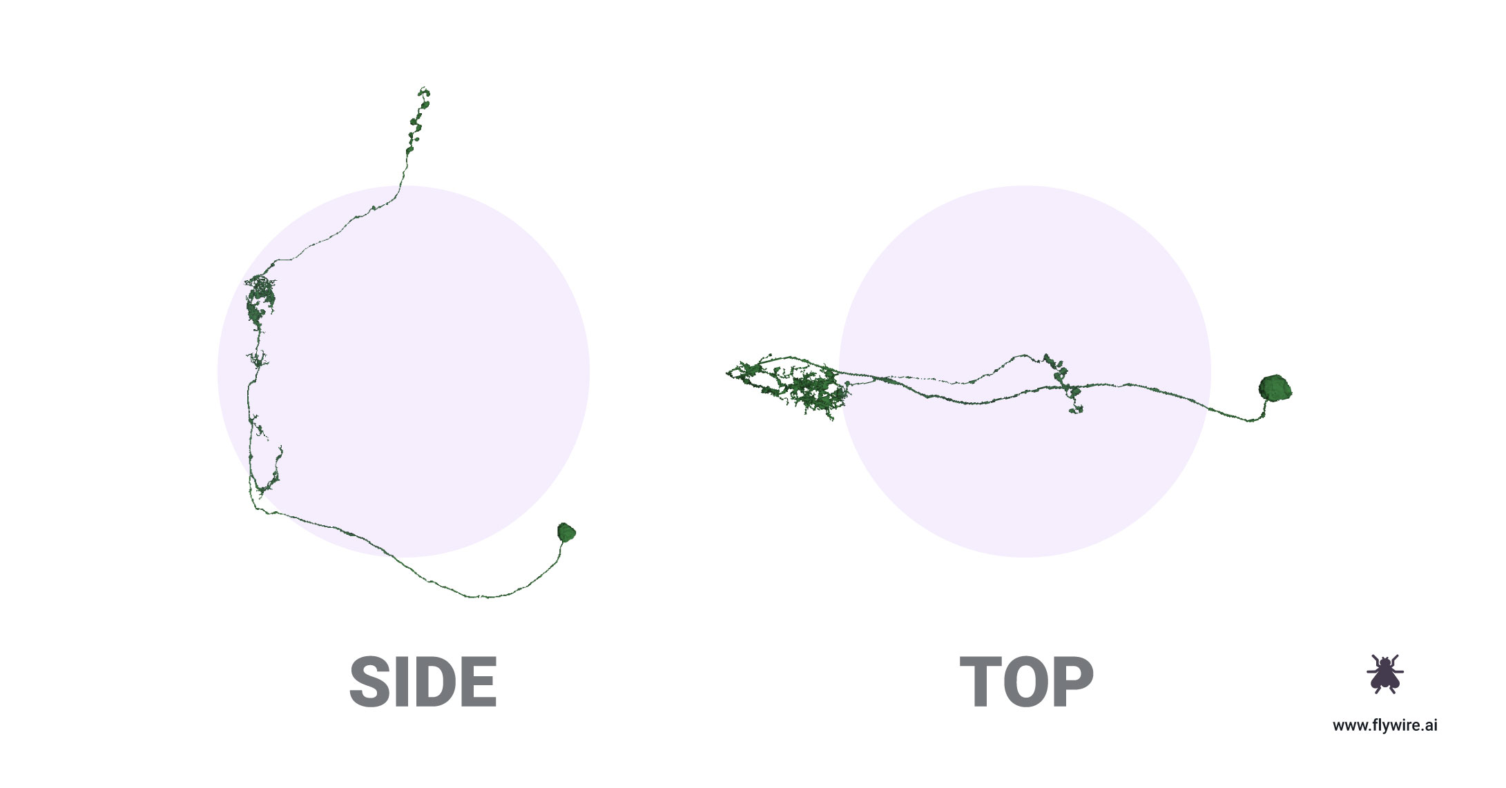
These neurons do something cool, probably.
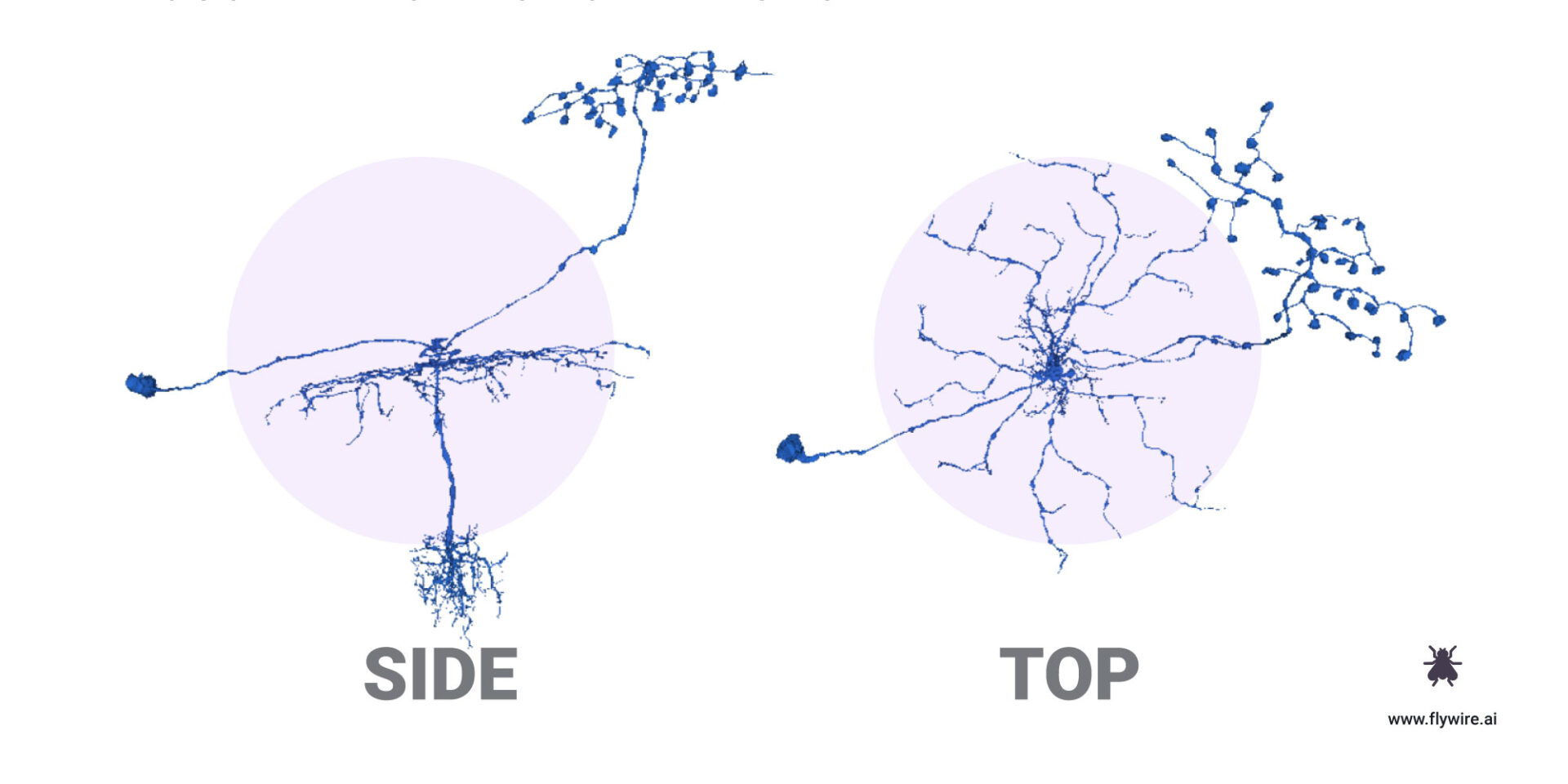
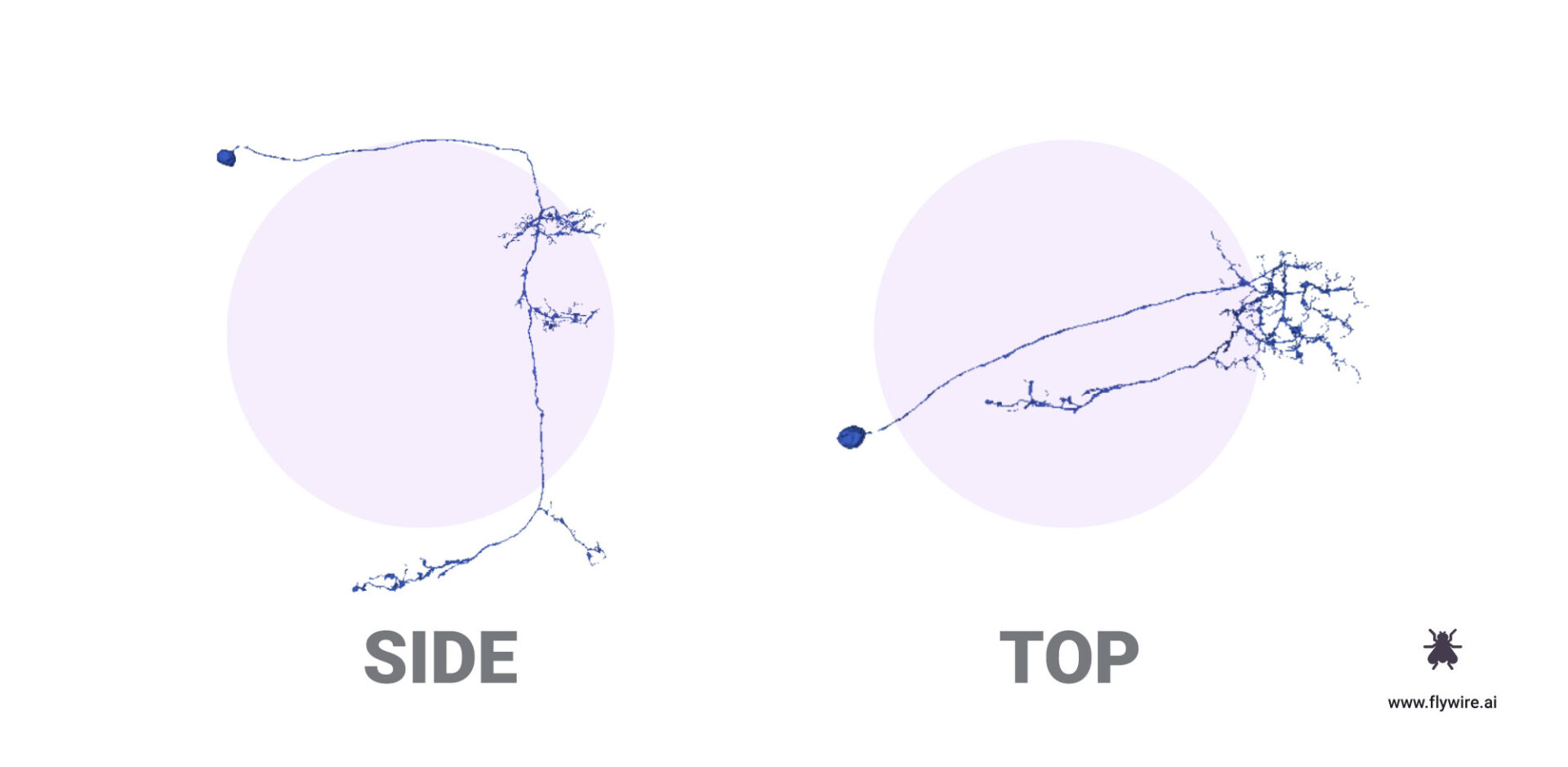
Distal Medulla (DM) Cells
FlyWire Link to example cells. Cells relative to neuropil.
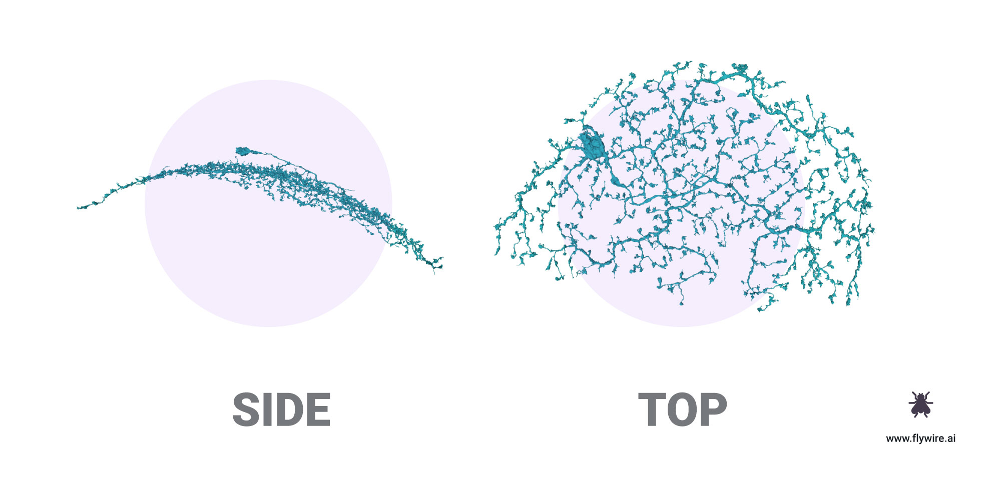
These canopy-esque cells spread branches into wide fields of photoreceptors. They may be motion sensitive and have a preferred directional stimuli, like ganglion cells of the retina.
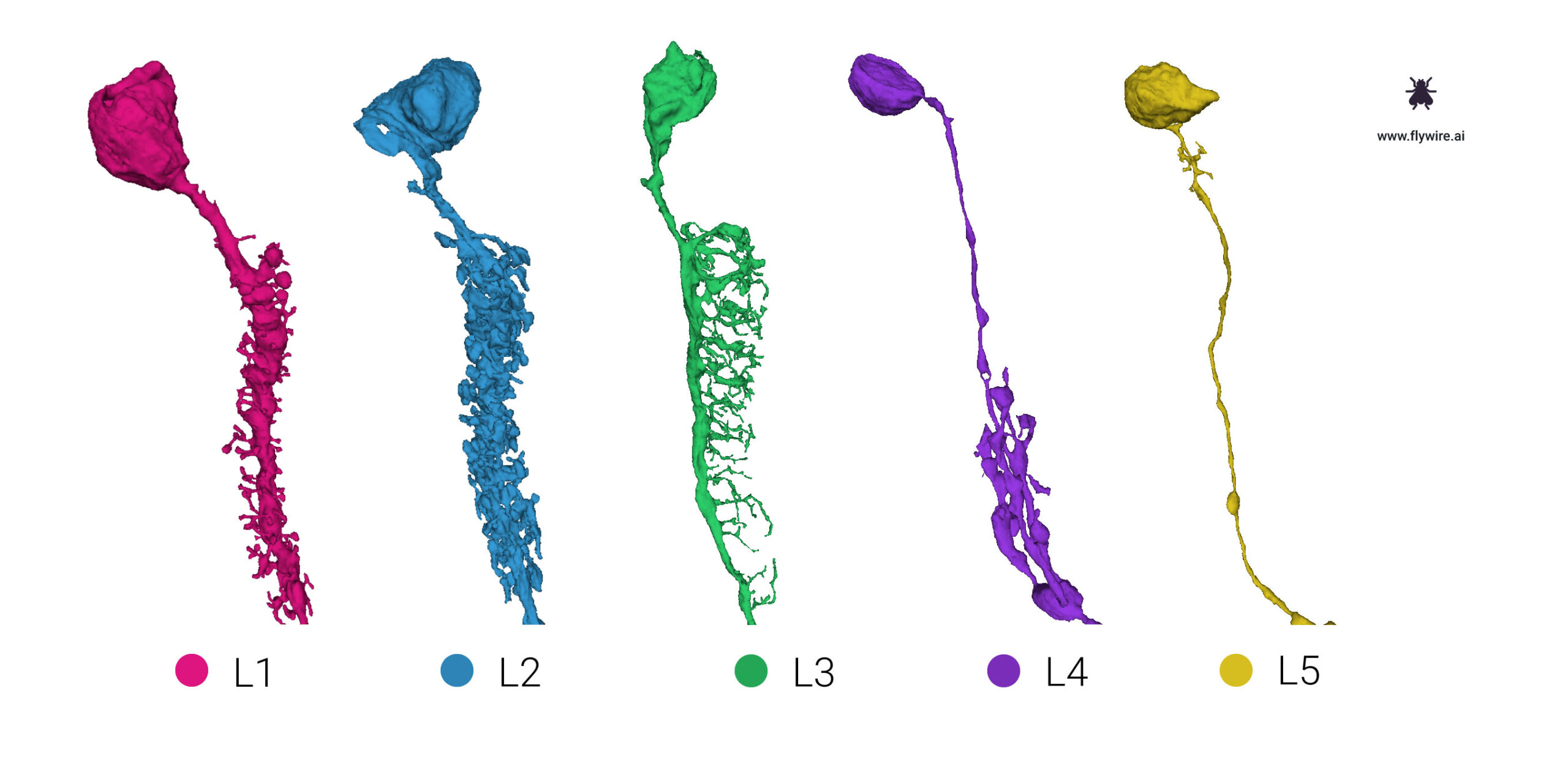
Kelp – Laminar Monopolar (L1-5)
FlyWire link to example cells.

These exotic neurons have somas located in the lamina and axons that reach into the Medulla. The number after L indicates where their branches stratify.
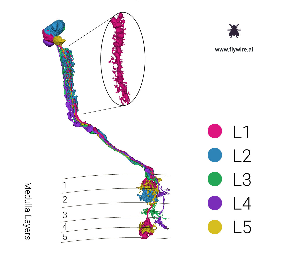
The necks of these neurons exhibits characteristic dendritic morphology which may aid in identifying cell types.
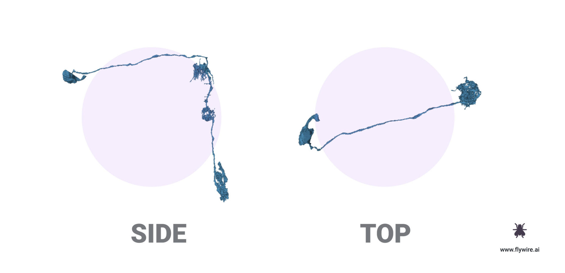
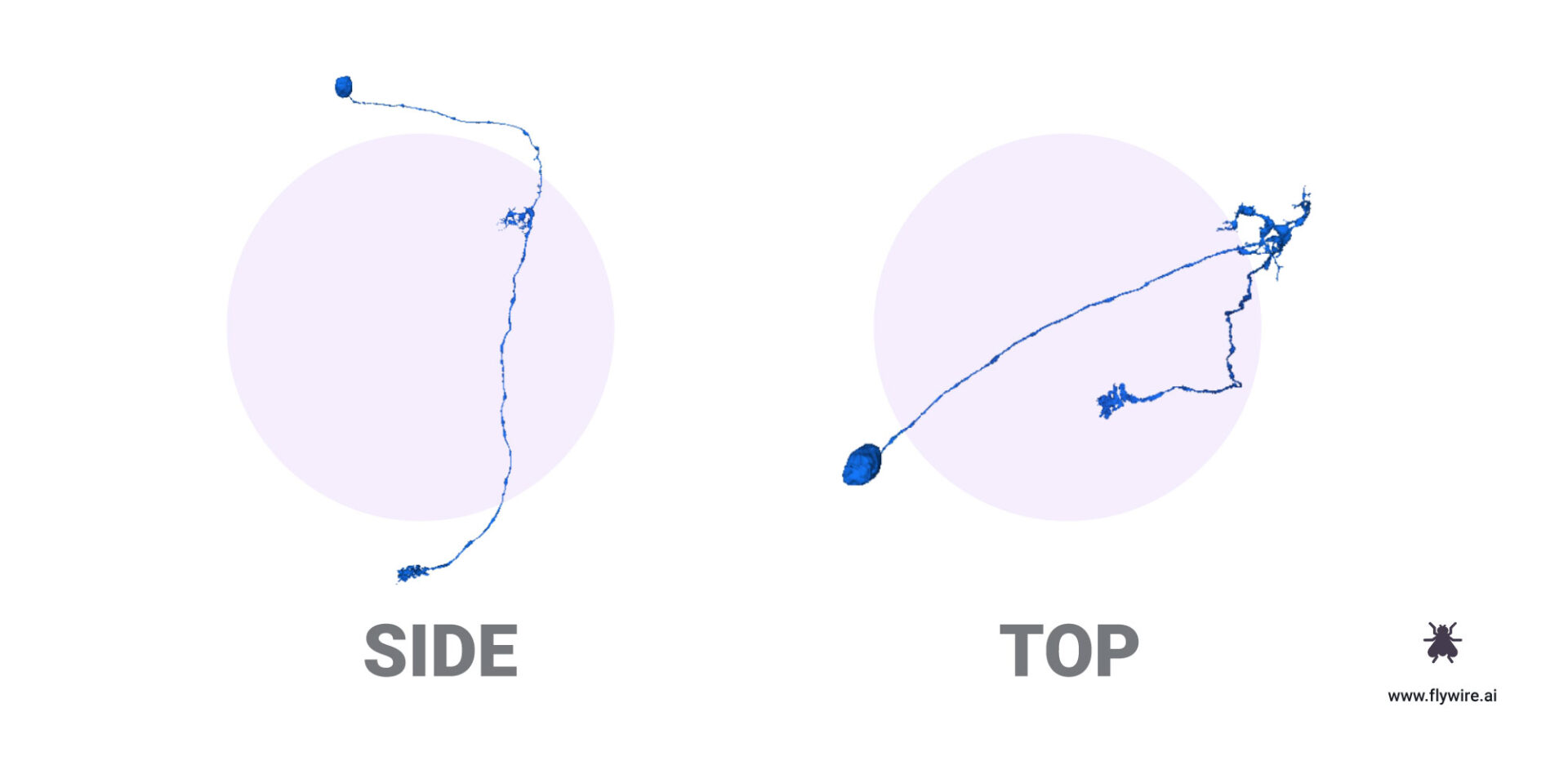
Laminar Wide Field Interneuron (LAWF)
FlyWire link to example cells.
These cells have their soma in the medulla and stratify into distinctive layers. Their dendrites can be found in the lamina while their axons reach into the medulla.
LAWf neurons with respect to the whole fly brain.
Spy Neurons – Medulla Intrinsic (MI)
FlyWire link to example MI neurons.
MI neurons connect within the medulla. Specifically, the distal (farther from central/closer to the eye) with the proximal (closer to central brain). Their somas are found in the medulla cortex.
Trans (T) Cells
FlyWire link to T cells. T cells relative to neuropil.

These “U-Turn” shaped cells send dendrites from their somas in the lobula up into the medulla, then their axons reach back down into the lobula.
These neurons are very numerous. T4 and T5 in particular. There are 4 different T4s and T5s in each column (a,b,c,d). With about 800 columns per optic lobe, that makes about 12,800 T neurons.
Trans-Medulla (TM)
FlyWire link to TM cells relative to neuropil.
Trans Medulla cells span the medulla.
Trans Medulla Y (TMY)
FlyWire link to TMY cells. TMY cells relative to neuropil.
The “Y” shape of TMY neurons allows these cells to make connections in two areas. These cells connect the distal medulla with lobula and lobula plate.
If you made it this far, bravo! We will continually update this page as new information is uncovered. You have finally reached the end of the optic column cell catalog and the beginning of a new era of connectomics. For Science!
-Amy
Thanks to Emil Kind for his extensive scientific advice on this post.
4 responses to “Optic Lobe Cell Catalog”
[…] excited to share that FlyWire Zone 2 is complete! Congratulations! Hundreds of neurons from the first 9 optical columns have been […]
[…] It’s always helpful to have a reference to compare your work against! Currently our production dataset for FlyWire players concerns the optic lobe. We’ve got examples of these types of cells here! […]
[…] It’s always helpful to have a reference to compare your work against! Currently our production dataset for FlyWire players concerns the optic lobe. We’ve got examples of these types of cells here! […]
If human eyes were proportionally as large as a fly’s, how would our vision and appearance change, resembling anime eyes? Regard Telkom University