
Calling all Flyers to a new quest! We’re on the hunt for unlabeled retinula cells, and we need your help to complete them!
These photoreceptors extend into the retina, whereas our dataset terminates at the lamina. Therefore, it will not be possible to fully complete these cells, though we can observe large enough sections for accurate labeling. Each may also be declared “complete” once the viewable portion of the cell has been proofread.
How it works
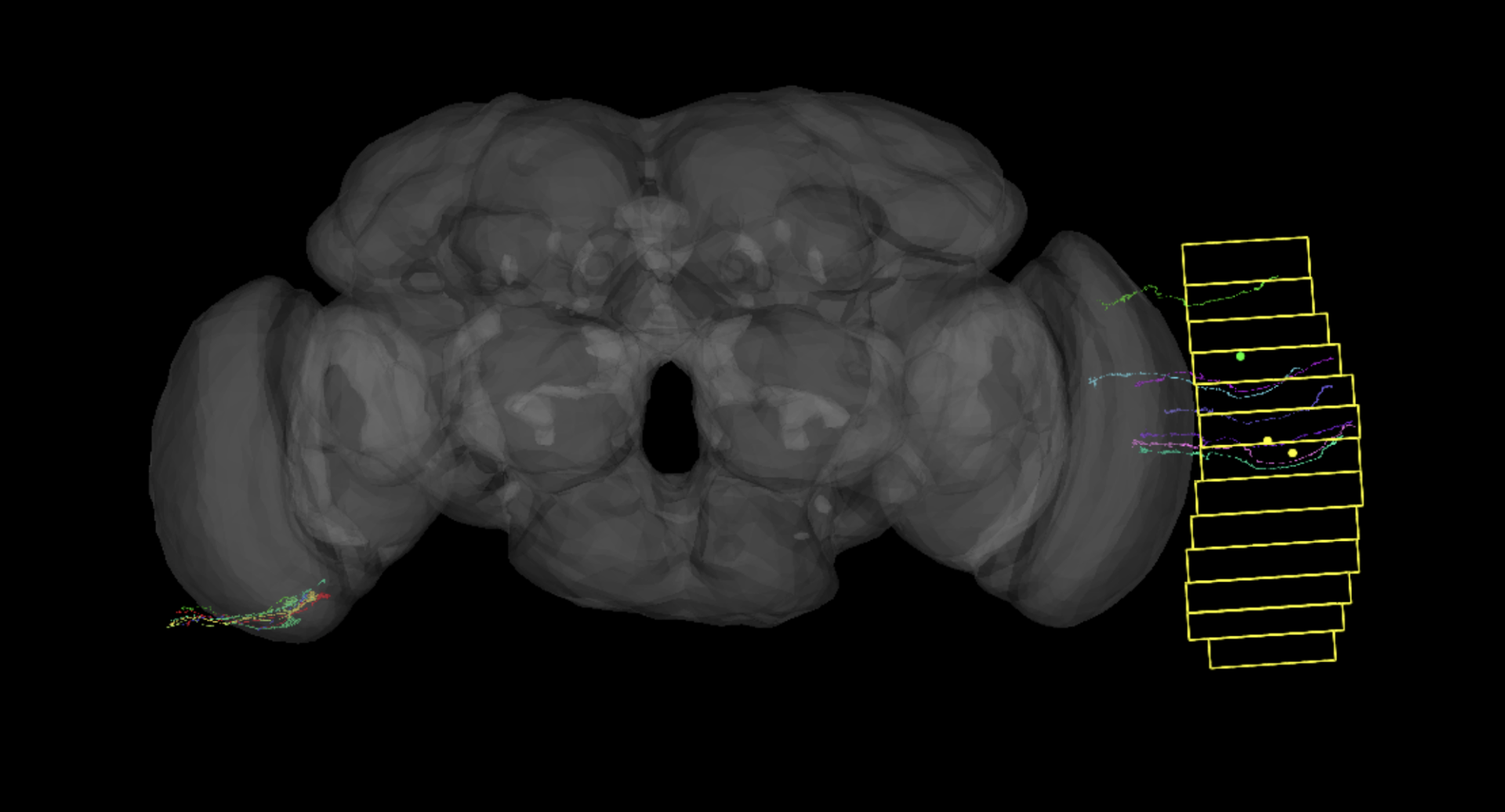
The quest will begin in a page from the FlyWire Q&A Log, which can be found here.
- Claim one of the available sections by entering your name in the “Username” section
- Mark the section’s status as “In Progress”
- Open the link
- Complete and label all unlabeled R cells in your section, which is indicated by a yellow bounding box
- Mark your section’s status as “Complete”
- If you need to abandon your section for an extended period of time, please mark the cell as “Need assistance,” so that another Flyer may take over for you
- Post to the FlyWire Forum when a section is completed
- The forum is also a great place to congratulate and encourage your fellow Flyers as we complete this collaborative quest!
Identifying + Labeling R cells
The axons of R 1-6 cells extend through the lamina, but also terminate within the lamina. They are recognizable by a dense, spiny axonal terminus.
R7 and R8 cells extend through the lamina, and then down into the medulla. R7 cells become knobby as they enter the medulla, then thin out, and then terminate with a club-like shape. R8 cells terminate slightly earlier and have a more uniform shape.
There are also R7 and R8 DRA (dorsal rim area) cells. With DRA cells both R7 and R8 terminate close to each other so they can be difficult to distinguish from one another. We recommend labeling them in pairs for best results.
We will also be labeling DRA cell types as “putative” since the R7 DRA and R8 DRA types can be difficult to distinguish from one another.
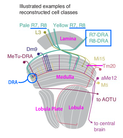
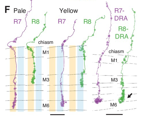
*Note: we are not currently distinguishing between pale and yellow R7/R8 cell types, so you can continue to label non-DRA types as “R7” or “R8.”
Please note that glial fragments tend to like to stick to these cell models, so you may have to do a good amount of pruning in order to free some of them.
Here are some examples of Retinula cells:
R7 + R8 (R7 are pink, R8 are green)
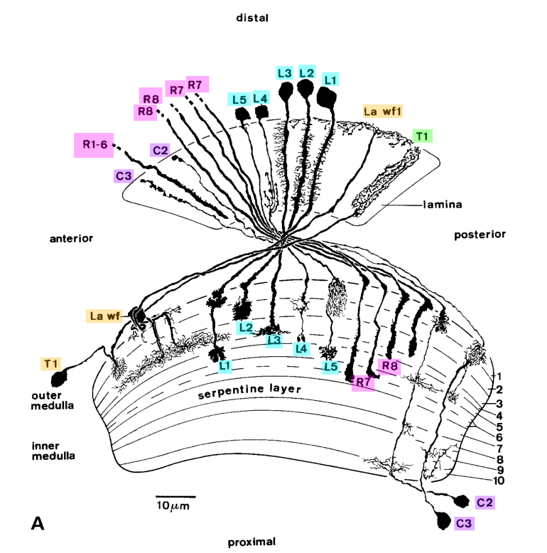
If you are having trouble with the distinction between R7 and R8 cells, you can simply label them as Retinula axons. We recommend the following labels depending on cell type:
R1-6 Label: Retinula axon 1-6; R1-6
R7 Label: Retinula axon 7; R7
R7 DRA Label: Retinula axon R7 dorsal rim area; putative R7-DRA
R8 Label: Retinula axon 8; R8
R8 DRA Label: Retinula axon R8 dorsal rim area; putative R8-DRA
R7 or R8 (unsure) Label: Retinula axon 7 or 8; R7/R8
Helpful tools
For easy labeling we suggest you enable @Krzysztof Kruk’s “Cell Identification Helper” addon which can be downloaded here.
If you have not used addons before, please see this blog post for instructions on how to install tampermonkey.