Big thanks to FlyWire team member Szi-chieh for putting together this guide for finding photoreceptors!
Introduction
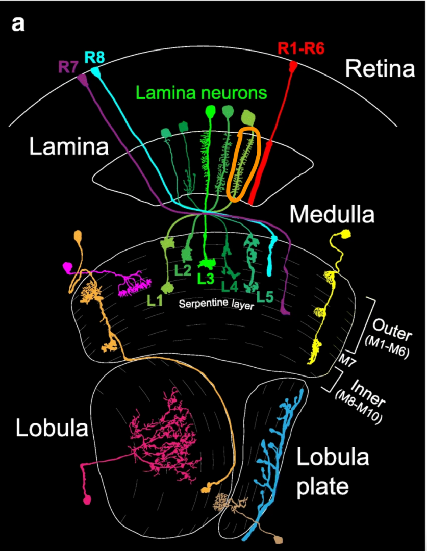
Drosophila has two chief motion detection pathways (reference):
- L1 → Mi1 (and C3) → T4
- L2 → Tm1 (Tm14 and Mi8) → T5
Since we have all Mi1 cells labeled in the left hemisphere, based on the motion detection pathways, we can work our way up using an individual Mi1 to locate its presynaptic partner L1. Then we can use the lamina part of the L1 to locate R1-6.

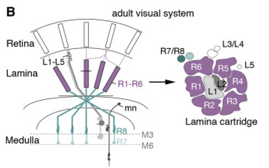
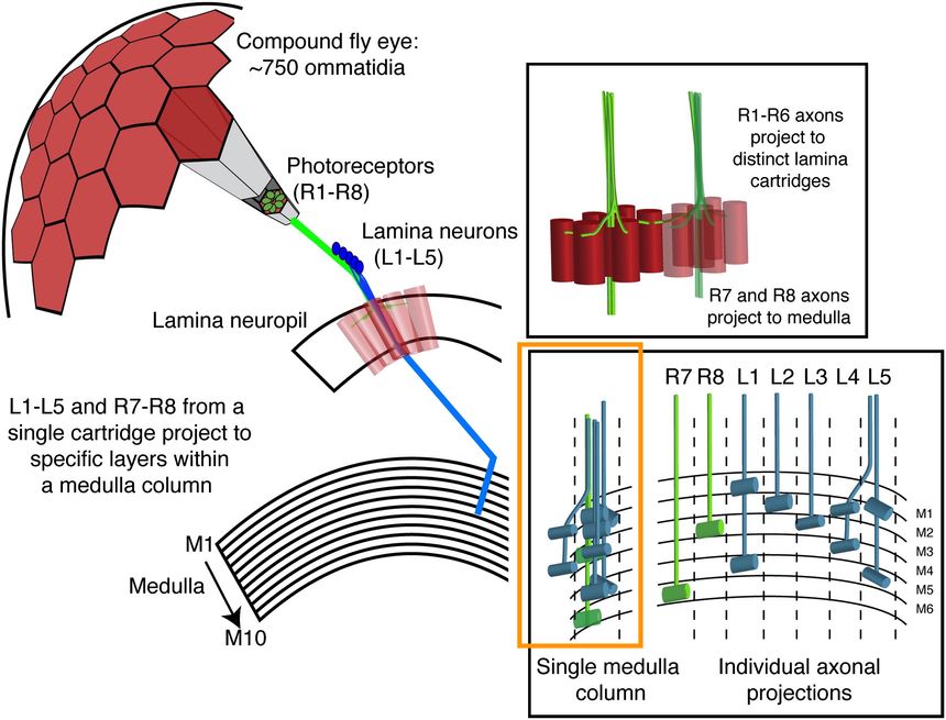
Furthermore, as the arbors of R7, R8 and L1 from a single lamina cartridge project to the same medulla column (yellow box from bellow image), we can use the medullar arbor of L1 to find R7 and R8 in our desired lamina cartridge.
How it works
1. Start with an Mi1
FlyWire link to 797 Mi1 cells in the left optic lobe (by KrzysztofKruk)
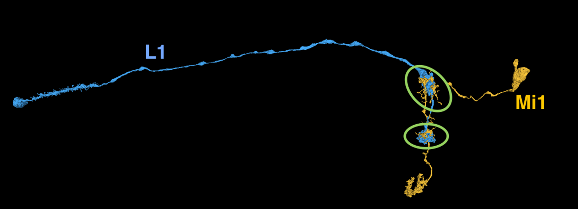
2. Use Mi1 to find L1
Mi1 synapses with L1 at these circled locations
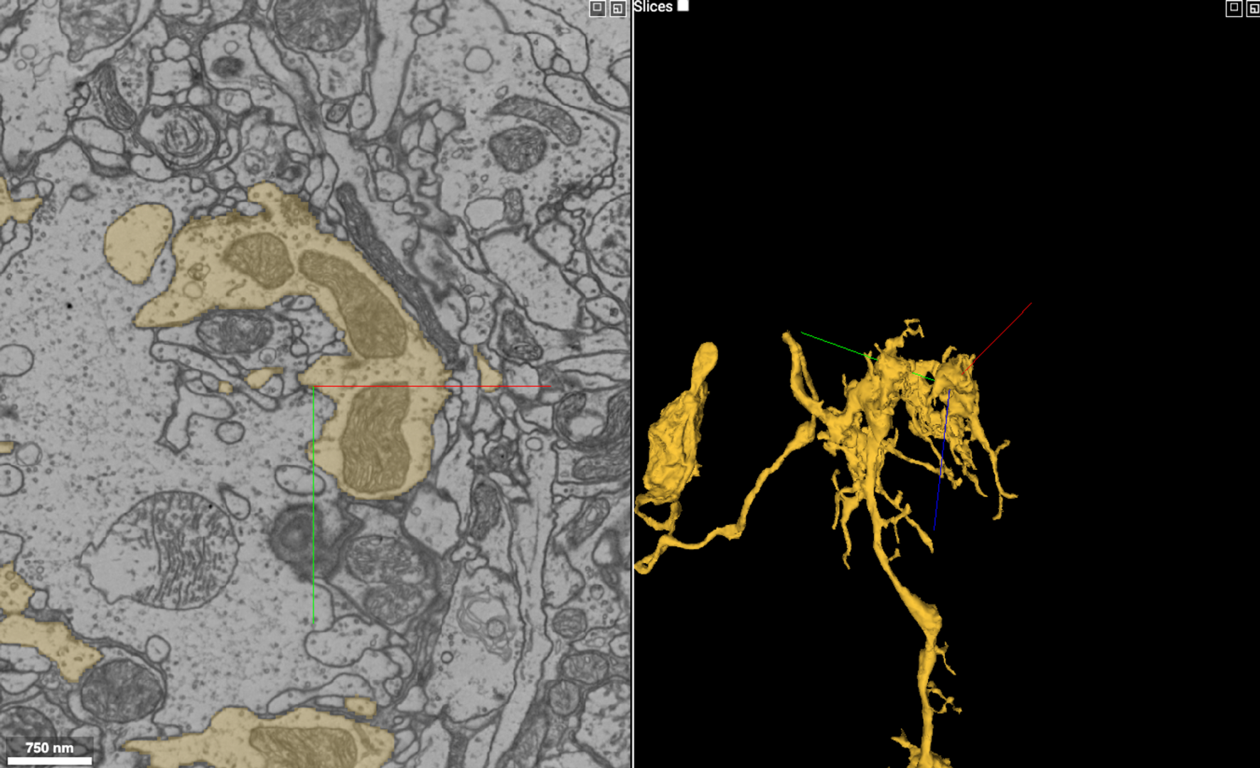
Find a twig based on the locations provided above. Once you find a synapse like the one shown at the crosshairs in the image below, select its presynaptic partner – usually the L1 cell. (To find out more about fly synapse ultrastructure, check out the fly_synapses_sunglab pdf file).
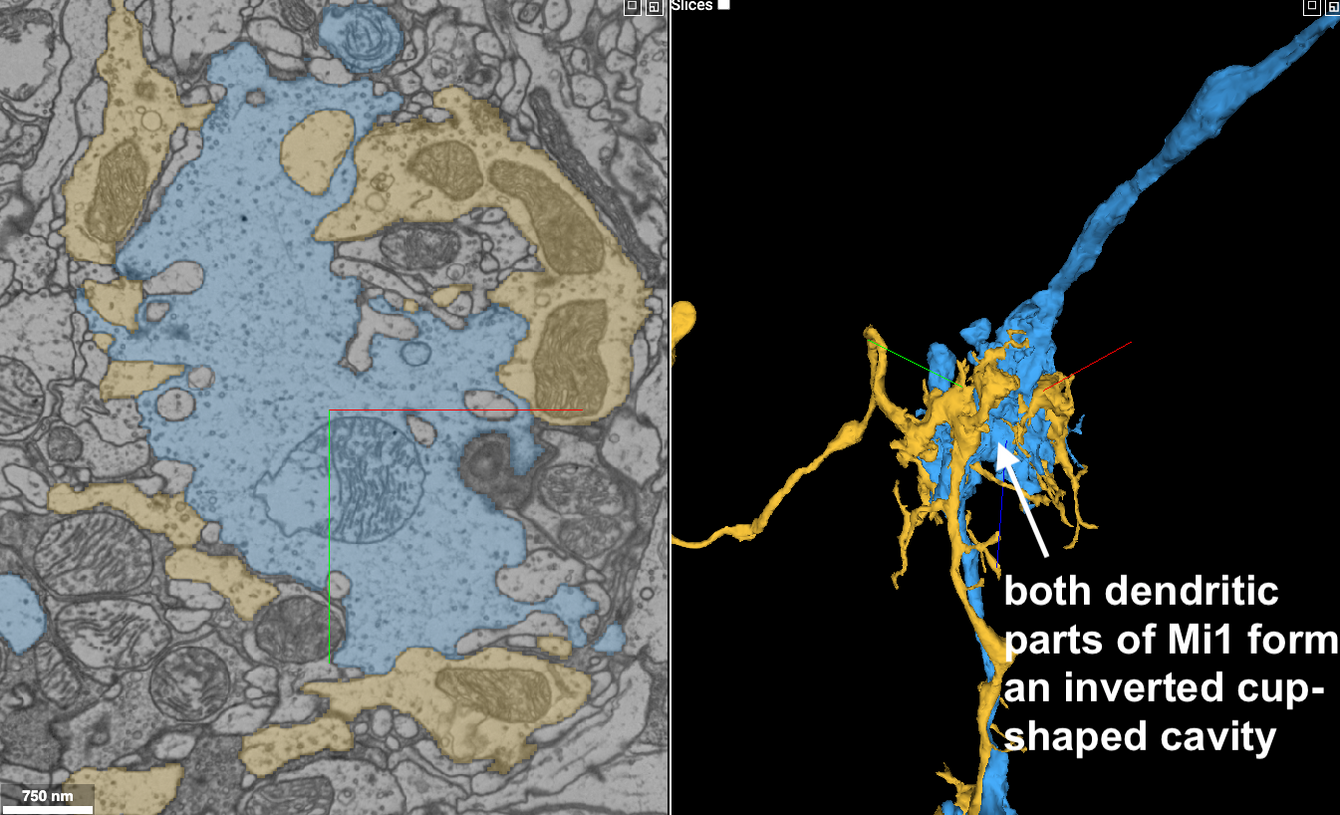
Sometimes you can also select the segment inside the “dendritic cavity” (which might also be the larger segment near the Mi1) to locate the L1.
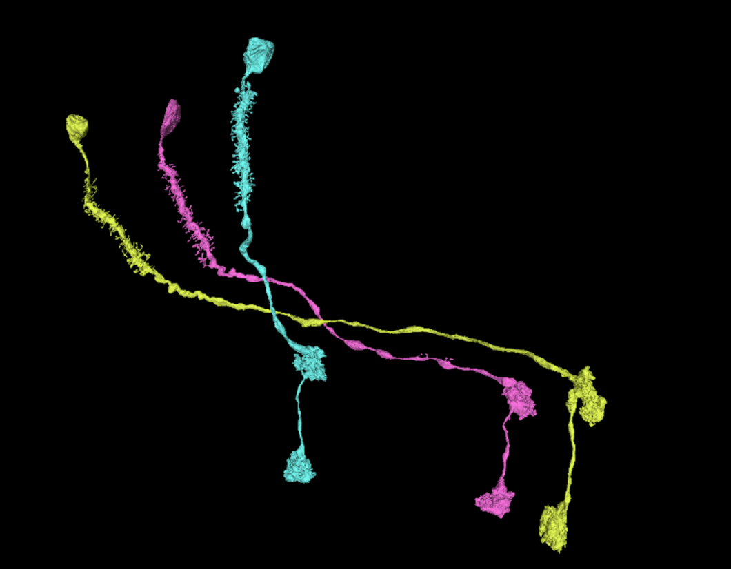
Visual of L1 cells in FlyWire:
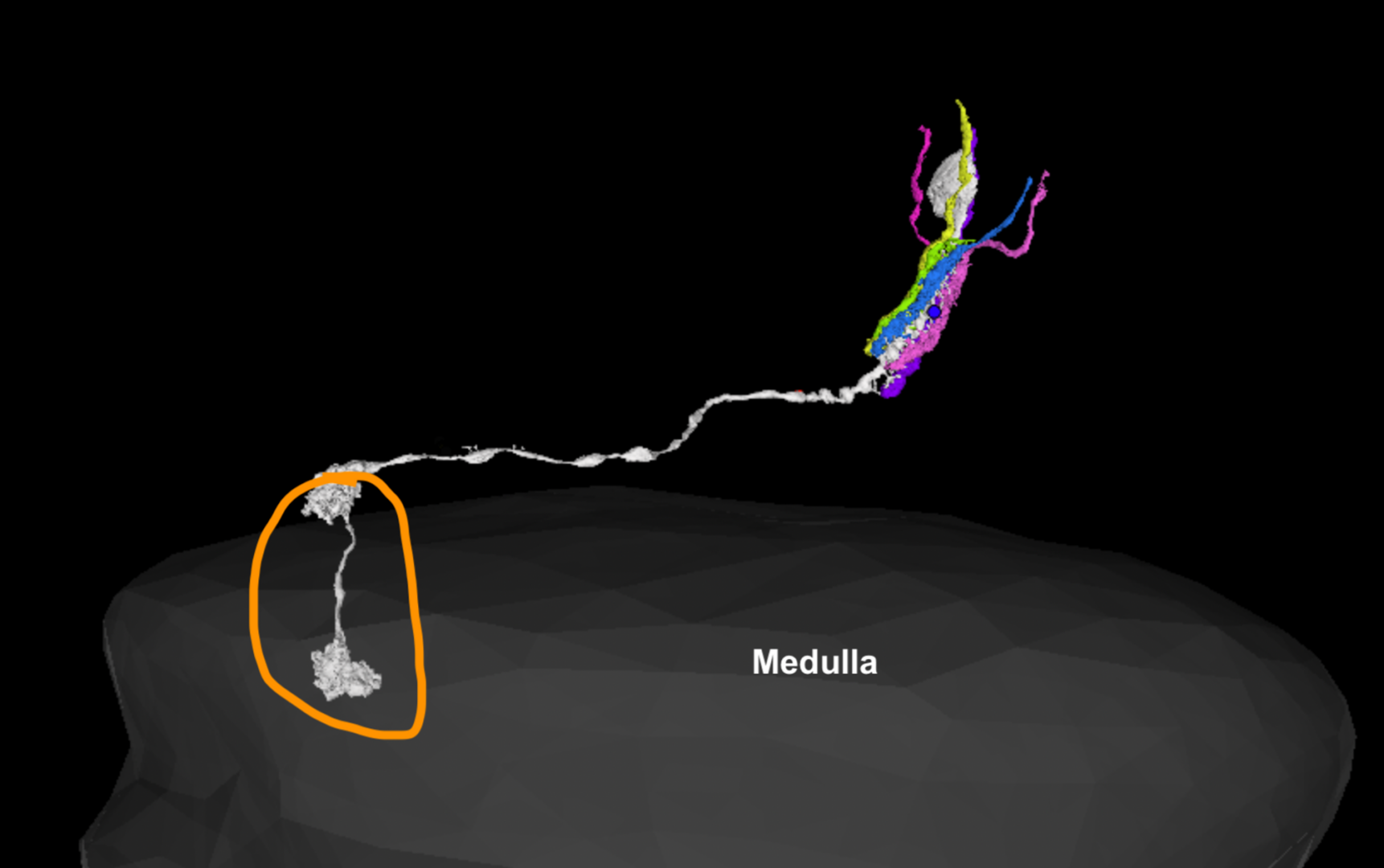
3. Using the lamina part of L1 to find photoreceptors R1-6
Go to the lamina part of the L1 (circled in yellow on the following image)
Use “s” on the keyboard to bring up the section plane. You may need to use “space” on the keyboard to toggle the layout so the section plane is a cross section through the L1 lamina region.
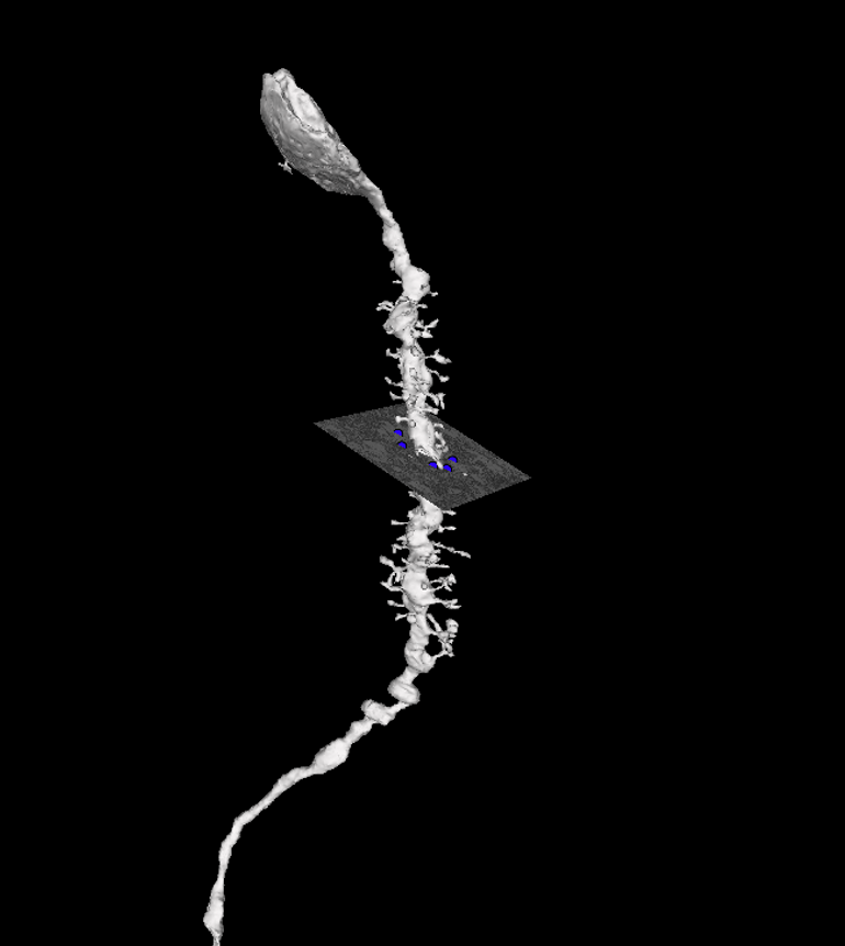
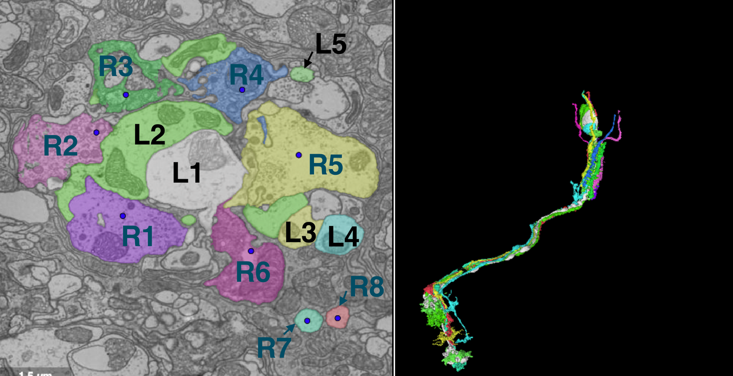
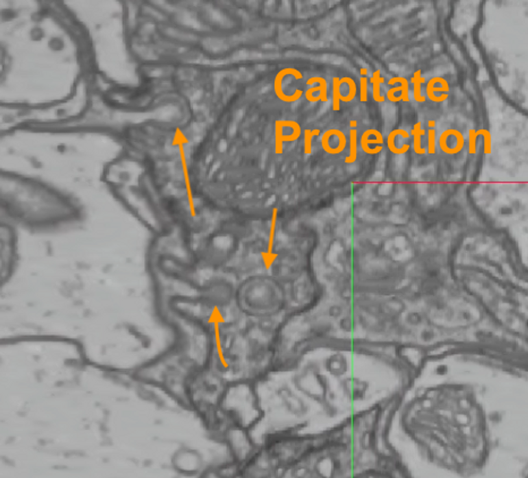
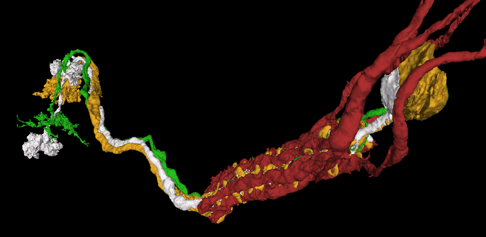
From the cross section, you can find R1-6 via an ultrastructure called capitate projection (glial processes inserted into R1-6). They have very distinctive double layer structure.
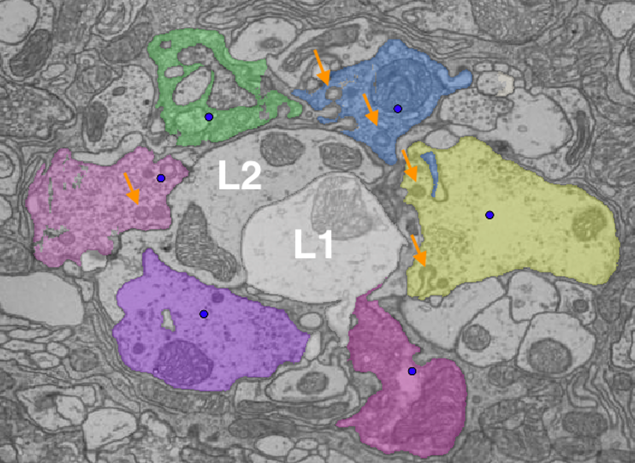
In a good cross section, L1 & L2 are in the center of a lamina cartridge and all R1-6 are circled around them very closely. (colored segments: R1-6; yellow arrows: capitate projection)
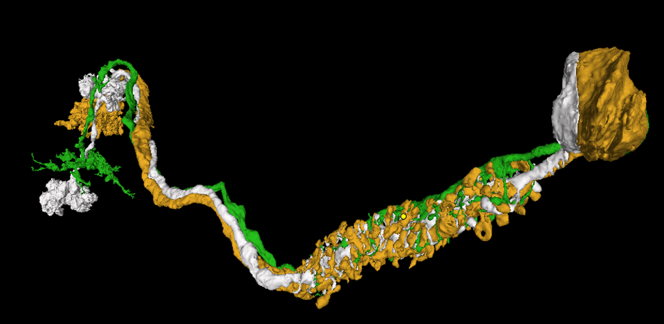
Advanced guide to find R1-6 in the same lamina cartridge
L1 and L2 have rich synapses from R1-6 and L3 is postsynaptic at a subset of them. Using the twigs from L1-L3, you can find the circumference of the R1-6 locations (see dashed blue line from the image). FlyWire link (206934, 55319, 2882)
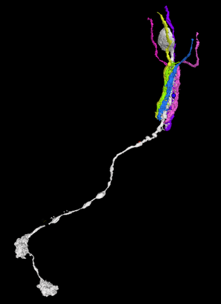
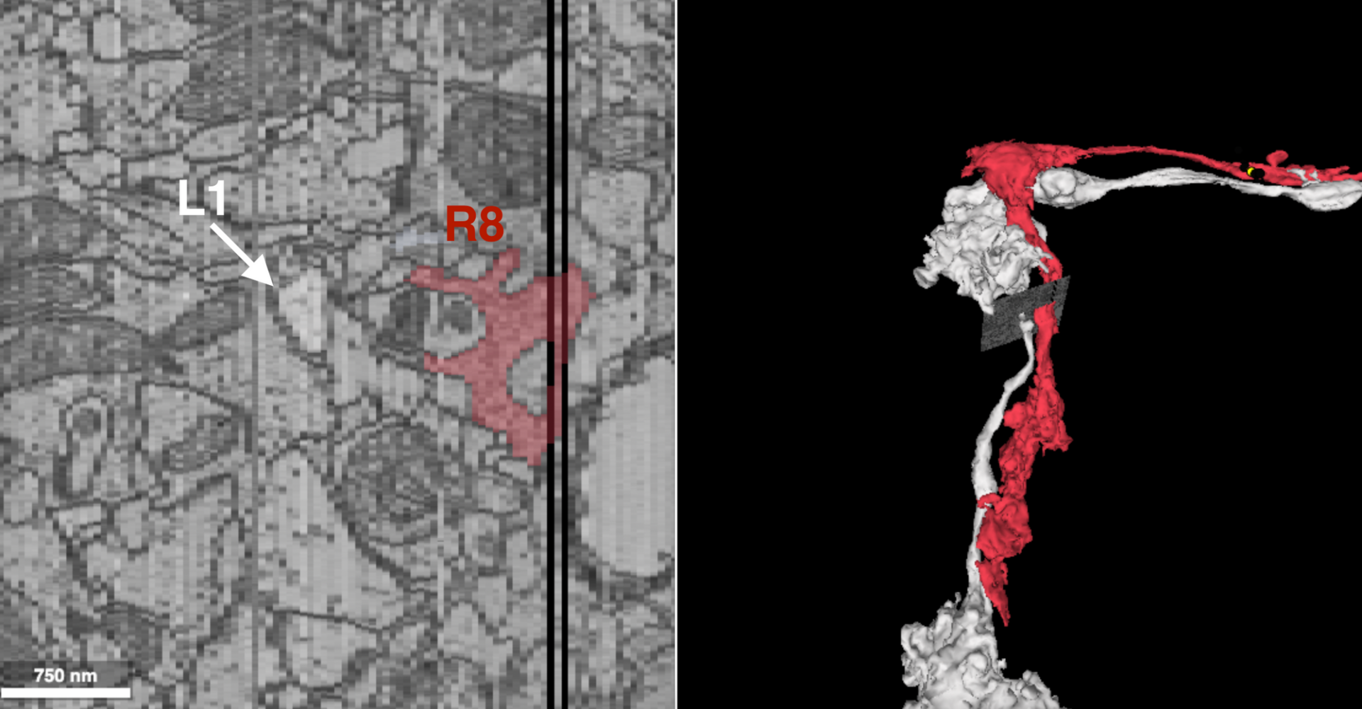
4. Using medulla part of L1 to find R7 & R8
L1, R7 and R8 in addition to other cells form a column in medulla, so we can use the medulla part of L1 to locate the R7 and R8 that are in the same cartridge of L1 & R1-6 (actually, you can find L2-L5 from this region, too!)
Use “s” from the keyboard again to find a good cross-section layout.
Click around to find cells that travels along with L1. The medullar arbors of L1-5 & R7-8 all travel together. Once you find either R7 or R8, then you can find its counterpart by locating a good cross-sectioned region from the R7/8 you find along the branch (does not necessarily need to be in the medulla.)
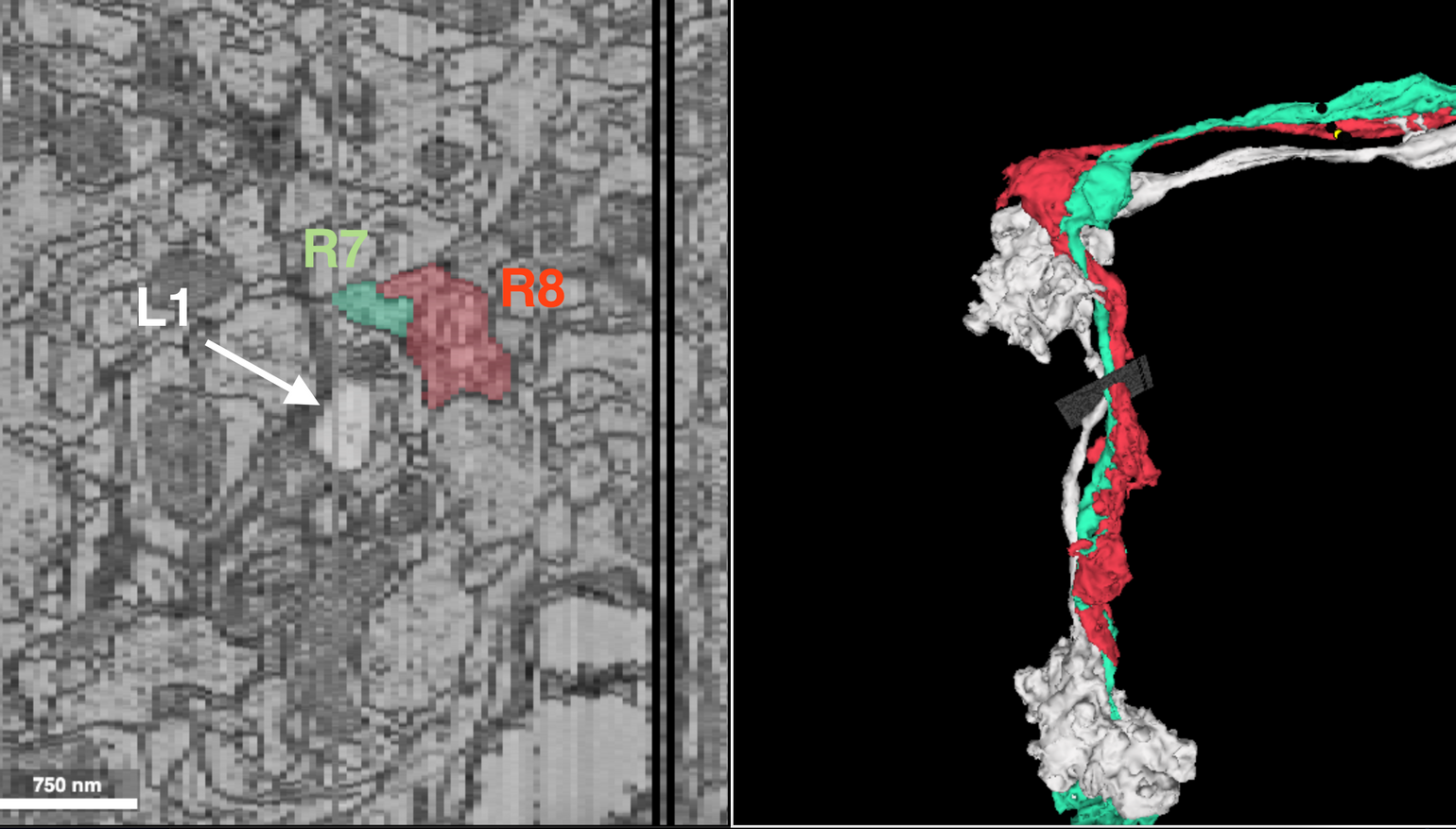
Now you have them! All R1-8 from a single cartridge/column. Congrats!
Illustration of the position of R1-6 and L1-5 in the lamina cartridge and FlyWire example